
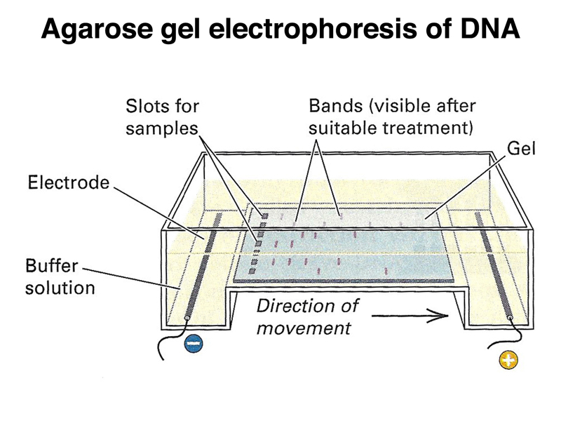
First we will see how a agarose gel electrophoresis looks like.

Image of an agarose gel electrophoresis apparatus
Basic Principle
Gel Electrophoresis is widely used technique for analysis of nucleic acid and proteins. It is mostly used to separate single and double stranded DNA molecules. As we know that, DNA has a negative charge due to the presence of a phosphate group, so the negatively charged DNA molecule migrates towards the anode (positive charge) when electric field is applied. The DNA then moves through the agarose gel according to their size and provide a sieving effect. The smaller DNA fragments moves farther away as compared to larger fragments. After the gel is allowed to run for a sufficient amount of time bands of DNA can be observed only after staining them with ethidium bromide.

The gel used in agarose gel electrophoresis is agarose which is a polysachharide extracted from sea weed. Agarose gel is linear type polymer that consist of a long chain of D and L galactose in an alternative manner. The structure of agarose gel is given below.

Structure of agarose gel
Choice of buffer
Gel electrophoresis requires a medium to run so the choice of buffer is important.
TAE buffer is best suited for gel elctrophoresis. Where T stand for Tris(strong base), A stands for Acetate(acid) and E stands for EDTA. The TAE buffer is slightly in alkaline range as under these circumstances DNA is protected from hydrolysis.Here EDTA is a chelating agent that binds with divalent cations specially magnesium(Mg2+) as in absence of magnesium ions the enzyme DNase is inactive which is responsible for degradation of DNA.
Loading buffer
The loading buffer is mixed with the DNA sample. The purpose of this buffer is to allow us to see the sample in gel as it contain a dye known as Bromophenol blue, and this buffer also contains glycerol which increases the density of the sample allowing them to sink in the wells properly.
Ethidium Bromide
As mentioned above ethidium bromide is used to stain the DNA as it binds with the DNA and gives fluorescence under microscope. Ethidium bromide is already present in the gel and intercalates between the stacked DNA.

Sample of DNA under UV light
DNA Ladder/ RNA Ladder/ Protein Ladder
A DNA ladder is the loaded in the first well of gel electrophoresis as it used as a reference to know the size of the given strand of the DNA.
Conclusion
So this is how our desired DNA is separated on the basis of its size or molecular weight. This technique is widely used in a biochemistry lab and should also be noted that this is not an alternative for purification of DNA as during this process our DNA gets denatured.
1 Comments
I am a co-author on the 3rd edition of "Basic Laboratory Methods for Biotechnology" to be published by CRC Press. I am trying to find who I can contact to receive permission to publish the diagram of agarose gel electrophoresis on your blog. Thanks for any information. Best regards,
ReplyDeleteJeanette